Prof. Dr. Alexandre Campos Moraes Amato
Professor of Vascular and Endovascular Surgery, UNISA
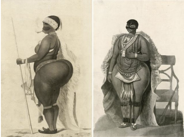
The shocking story of an African woman of the Khoisan people who became a circus attraction owing to ignorance and prejudice. On 29 December 1815, 125 years before lipedema was described by Allen and Hines1, Saartjie Baartman (Figure 1) died after spending years being exhibited at European fairs as a “bizarre human phenomenon.” Her brain, skeleton, and sexual organs continued to be displayed in a Paris museum until 1974. Her remains were only returned to Africa in 2002, after France agreed to a request made by Nelson Mandela. She had apparently been taken to Europe under false promises by a British physician. She was given the stage name “The Hottentot Venus” (now considered an offensive term) and turned into a circus attraction in London and Paris, where crowds gawked at her buttocks. For an extra fee, her exhibitors allowed visitors to touch her buttocks, whose unusual volume (steatopygia) struck the European of the time as strange and disturbing.
In late 1814, Saartjie was sold to a Frenchman, an animal tamer who saw in her an opportunity for easy enrichment. Regarding her as having been acquired as a prostitute or a slave, her new owner kept her under far harsher conditions. She was exhibited in Paris, forced to display herself completely nude, which violated her vow never to show her genitals. Napoleon Bonaparte’s celebrations in early 1815 included nighttime parties where the many drunken visitors amused themselves by groping the body of the defenseless woman.
She was then exposed to crowds who mocked her. She was the target of caricatures, but she also drew the interest of scientists and painters. French anatomists and other naturalists visited her, and she became the subject of numerous scientific illustrations at the Jardin du Roi. Her body was thoroughly examined and measured, with records made of the size of her buttocks, clitoris, labia, and nipples for zoological and scientific museums and institutes.
The dissection of her body was carried out and published by the French anatomist Henri Marie Ducrotay de Blainville (1816) and republished by the French naturalist Georges Cuvier in Mémoires du Museum d’Histoire Naturelle (1817). Cuvier, who had known Baartman, noted in his monograph that his object of study was an intelligent woman with an excellent memory, especially for faces. In addition to her native language, she spoke Dutch fluently, had passable English, and some knowledge of French. He describes her shoulders and back as “graceful,” with “slender arms,” and her hands and feet as “charming” and “pretty.” He adds that she was skilled with the mouth harp, danced according to the traditions of her country, and had a cheerful personality.
Diseases that lack specific markers, gold-standard tests, or imaging studies are often discredited. Other diseases whose diagnosis was always clinical have come to require imaging studies in order to prove their existence to the health system. As a result, subsidiary tests have often come to be regarded as definitive, while the importance of history-taking and physical examination is relegated to the background. Diseases that present wide clinical variation and lack definitive subsidiary tests are forgotten and are slow to be recognized. For the generalist, the difficulty in clinical diagnosis lies in recognizing conditions that do not fit into the familiar, already-known disease categories, and lipedema is one of them. As mentioned earlier, lipedema was first described in 1940 by Doctors Edgar Van Nuys Allen, a cardiovascular surgeon known for the Allen test, and Edgar Alphonso Hines Jr. at the Mayo Clinic2,3 in the Vascular Clinics session, and today it names the Allen-Hines syndrome4. Since then, lipedema has been characterized as an abnormal deposition of fat in the buttocks and legs bilaterally, which may be accompanied by orthostatic edema2,3.
The pathophysiology and epidemiology of lipedema are poorly understood, and for this reason it is not included in the basic academic medical curriculum, and often not even in the specialized vascular curriculum. It is therefore frequently confused with more common conditions such as obesity and lymphedema5,6. Although it was initially described in the United States, the most recent advances have emerged in Europe, chiefly through the work of professors Michael Földi and Etelka Földi7 in Germany, which has only recently gained greater recognition in the United States8 and in Brazil9–12. Awareness of lipedema among the lay public has increased in recent years, partly owing to the proliferation of internet content, online groups, and the beginning of mainstream media recognition of the condition, but the stigma of the disease still exists within the medical community.
The disproportionate fat distribution typical of lipedema has also been divided into five types, according to the area affected, with some patients classifiable into more than one type13.
- Type I: pelvis, buttocks, and hips (Figure 1).
- Type II: buttocks to the knees, with a fatty pad folded around the inner side of the knee.
- Type III: buttocks to the ankles.
- Type IV: arms.
- Type V: lower leg.
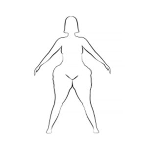
Considering current knowledge of lipedema, the anatomical description, and the images of the period, we can assess the history of Saartjie Baartman and conclude that she probably had the type I form of the disease known today as lipedema. Still in the realm of speculation, possibly at an advanced evolutionary stage, between stages 3 and 4 (lipolymphedema) of the disease.
Ignorance of the disease made her a target of caricatures and prejudice, and today she is a symbol of Western exploitation of Africans and of racism. But I believe that, beyond this, she is also an extreme example of the negative impact that ignorance of a disfiguring disease can have on a person. The period in which this took place also shows that lipedema is not a new disease. And the recognition of this bodily characteristic of Saartjie as prevalent among her people of origin also points to the genetic features of the disease, probably aggravated by the environmental factors to which that people were subjected.
Today we see lipedema in worldwide distribution, with lower prevalence among Eastern peoples.
May the suffering life story of Saartjie Baartman serve as an example to help us avoid prejudice and the stigmatization of bodily structure today. We must learn from the mistakes of the past and commit to doing better.
Bibliography
- Al-Ghadban, S., L. Teeler, M. & A. Bunnell, B. Estrogen as a Contributing Factor to the Development of Lipedema. in Physiology and Disorders of Adipose Tissue [Working Title] 32, 137–144 (IntechOpen, 2021).
- Allen, E. V, Hines, E. A. & Hines, E. A. Lipedema of the legs: a syndrome characterized by fat legs and orthostatic edema. Proc Staff Meet Mayo Clin. 15, 184–187 (1940).
- Wold, L., Hines, E. A. & Allen, E. V. Lipedema of the legs: a syndrome characterized by fat legs and edema. Ann Intern Med 34, 1243–1250 (1951).
- Whonamedit – dictionary of medical eponyms.pdf. Allen-Hines syndrome
- Fife, C. E., Maus, E. A. & Carter, M. J. Lipedema: a frequently misdiagnosed and misunderstood fatty deposition syndrome. Adv Ski. Wound Care 23, 81–84 (2010).
- Beninson, J. & Edelglass, J. W. Lipedema – the non-lymphatic masquerader. Angiology 35, 506–510 (1984).
- Asmussen, P. D. et al. Földi's textbook of lymphology for physicians and lymphedema therapists. (Elsevier Urban & Fischer, 2012).
- Schmeller, W. & Meier-Vollrath, I. Tumescent liposuction: a new and successful therapy for lipedema. J Cutan Med Surg 10, 7–10 (2006).
- Amato, A. C. M., Amato, F. C. M., Benitti, D. A. & Amato, L. G. L. Criação de questionário e modelo de rastreamento de lipedema. J. Vasc. Bras. 19, 1–7 (2020).
- Amato, A. C. M., Markus, D. V. & Santos, R. V. dos. Lipedema associado a obesidade, linfedema e insuficiência venosa: relato de um caso. Diagnóstico e Trat. 25, 4–8 (2020).
- Amato, A. C. M., Amato, F. C. M., Benitti, D. A. & Santos, R. V. dos. Tradução adaptação cultural e validação do questionário de avaliação sintomática do lipedema (QuASiL). J. Vasc. Bras. 7301, 1–8 (2020).
- Amato, A. C. M. Is Lipedema a Unique Entity? EC Clin. Med. Cases Reports 2, 1–7 (2020).
- Szél, E., Kemény, L., Groma, G. & Szolnoky, G. Pathophysiological dilemmas of lipedema. Med Hypotheses 83, 599–606 (2014).